
GUMBORO or IBD
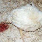
(Infectious Bursal Disease) is an acute, highly contagious viral infection in chickens manifested by inflammation and subsequent atrophy of the bursa of Fabricius, various degrees of nephroso-nephritis and immunosuppression. Clinically the disease is seen only in chickens older than 3 weeks. The feathers around the vent are usually stained with faeces containing plenty of urates.
The period of most apparent clinical symptoms and high death rate is at the age of 3 – 6 weeks. Gumboro/IBD could however be observed as long as chickens have a functioning bursa (up to the age of 16 weeks). In chickens younger than 3 weeks, IBD could be subclinical, but injured bursa leads to immunosuppression. Also, diarrhoea, anorexia, depression, ruffled feathers, especially in the region of the head and the neck are present.
A natural Gumboro/IBD infection is mostly observed in chickens The dead bodies are dehydrated, often with haemorrhages in the pectoral, thigh and abdominal muscles.
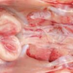
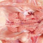
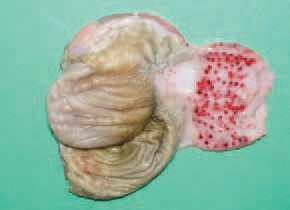
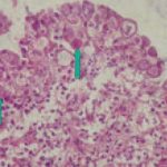
The Gumboro/IBD virus belongs to the Birnaviridae family of RNA viruses. Two serotypes are known to exist, but only serotype 1 is pathogenic. The virus is highly resistant to most disinfectants and environmental conditions. In contaminated premises, it could persist for months and in water, forage and faeces for weeks. The incubation period is short and the first symptoms appear 2-3 days after infection. The lesions in the bursa of Fabricius are progressive. In the beginning, the bursa is enlarged, oedematous and covered with a gelatinous transudate.
.GUMBORO/IBD virus has a lymphocidic effect and the most severe injuries are in the lymph follicles of the bursa of Fabricius. Most commonly, IBD begins as a serous bursitis. .IBD virus has a lymphocidic effect and the most severe injuries are in the lymph follicles of the bursa of Fabricius. Most commonly, IBD begins as a serous bursitis.
IBD lesions undergo various stages of serous haemorrhagic to severe haemorrhagic inflammation. The morbidity rate is very high and could reach 100%, whereas the mortality rate: 20 – 30%. The course of the disease is 5-7 days and the peak mortality occurs in the middle of this period.
In some cases, the bursa is filled with coagulated fibrinous exudate that usually forms casts with the shape of mucosal folds. In birds surviving the acute stage of the disease, the bursa is progressively atrophying. Microscopically, an atrophy of follicles into the bursa is observed secondary to inflammatory and dystrophic necrobiotic alterations. The kidneys are affected by a severe urate diathesis. In an acute outbreak and manifestation of the typical clinical signs, the diagnostics is not difficult. The diagnosis could be confirmed by detection of typical gross lesions throughout a patho-anatomical study. IBD should be differentiated from IBH (inclusion body hepatitis.
TREATMENT :
G1 GUMBORON :A unique combination of homoeopathic medicines highly effective against
Gumboro disease both prophylactically and therapeutically.
it is highly recommended to used as preventive
when it is used during disease for treatment G1 GUMBORON rapidly controls mortality and minimizes losses.
PREVENTIVE DOSE : 1ML / 2 LITER of water (One to three times a week for 4 weeks according to the outbreak severity in vicinity).
IN MILD DISEASE : 1ML / 1 LITER of water
IN SEVERE OUTBREAT: 2ML / 1 LITER of water
Available in packing of
500 ml and 1 liter
ND (Newcastle disease)
The Newcastle disease (ND) is a highly contagious disease in many species of domestic, exotic and wild birds that, depending on its tropism, is characterized by marked variations in morbidity, death rate, symptoms and lesions. The clinico-morphological signs possess a distinct viscerotropic or neurotropic character. In the viscerotropic form, haemorrhagic diphtheritic lesions of the entire alimentary tract, from the beak to the vent, are present. The haemorrhages of gizzard epithelium are remarkable. The mucous coat is oedematous, covered with thick mucus and mottled with haemorrhages varying from single to multiple, sometimes gathered at the boundaries with the gizzard or the oesophagus
Typical for this form are the haemorrhagic necrotic and focal diphtheroid lesions affecting the mucosa of the buccal cavity , the stomach and the intestines. The disease is generally prevalent in hens,peacocks, exotic or wild birds. Birds at any age are susceptible. It is caused by a paramyxovirus. Depending on their pathogenicity, the numerous known strains are classified as lentogenic, meso-genic and velogenic
A frequent finding is the enlargement and haemorrhages of caecal tonsils and haemorrhagic cloacitis. Usually, these lesions begin from the lymphoid tissue of the mucous coat. Virus-containing excreta of infected birds, that contaminate the forage, water and the environment, are the source of infection. The infection is transmitted mainly by an oral route, the airborne or contact transmission being more infrequent. The virus, contained in incubated eggs, results in embryo’s death and then perishes. There is no permanent carriership of the virus. An important factor in the trans¬mission of velogenic viruses could be the exotic birds and fighting cocks. The death rate could arrive at 70-100%
The neurotropic form of the disease is clinically manifested by ataxia, opisthotonus, torticolis, paresis and paralysis of legs. This form is frequently accompanied by respiratory symptoms. Histo-pathologically, the picture of nonpurulent lymphocytic encephalomyelitis is observed.
The lesions of paramyxovirosis in pigeons are entirely identical. On the basis of the history and the clinico-morphological signs, a tentative diagnosis could be made, but its laboratory confirmation is mandatory. ND should be distinguished by avian influenza, fowl cholera etc
TREATMENT :
G4 ND RON :A unique combination of homoeopathic medicines highly effective against Newcastle disease both prophylactically and therapeutically.
it is highly recommended to used as preventive
when it is used during disease for treatment G4 NDRON rapidly controls mortality and minimizes losses.
PREVENTIVE DOSE : 1ML / 2 LITER of water (One to three times a week for 4 weeks according to the outbreak severity in vicinity).
IN MILD DISEASE : 1ML / 1 LITER of water
IN SEVERE OUTBREAT: 2ML / 1 LITER of water
Available in packing of
500 ml and 1 liter
INFECTIOUS BRONCHITIS (IB)
In chickens up to the age of 4 weeks, IB is manifested by severe respiratory signs (sneezing, coughing, and rales). Rhinites and conjunctivites, depression and crowding around heat sources are observed. The death rate could reach 100%. The mortality in young chickens is usually insignificant provided that a secondary infection with a different agent is not occurring. In such cases, there is a moderate to severe inflammatory cell infiltration of upper respiratory tract mucosa, resulting In thickened and more compact mucosa. In layer hens infected with the IB virus, oophorites and dystrophic necrobiotic lesions affecting primarily the middle and the last thirds of oviduct’s mucous coat are observed. The consequences are drop in egg production, appearance and increase in the number of de¬formed and pigmentless eggs or eggs with soft shells and watery egg white.
The nephrotropic strains of the IB virus cause severe inflammatory and dystrophic necrobiotic damages of kidneys: urolithiasis, interstitial nephritis, haemorrhages that considerably increase the death rate. Under natural conditions, only hens are infected. Non-immune birds of all ages are susceptible. The disease is even seen in vaccinated flocks. The diagnostic method PCR is used for rapid identification of IB virus serotypes. IB should be distinguished from other acute respiratory disease as ND, laryngotracheitis and infectious coryza.
TREATMENT :
IB FORTE :A unique combination of homoeopathic medicines highly effective against
Infectious Bronchitis disease both prophylactically and therapeutically.
it is highly recommended to used as preventive as it reduces chances of outbreak in flocks.
when it is used during disease for treatment IB FORTE rapidly controls mortality and minimizes losses.
PREVENTIVE DOSE : 1ML / 2 LITER of water (One to three times a week for 4 weeks according to the outbreak severity in vicinity).
IN MILD DISEASE : 1ML / 1 LITER of water
IN SEVERE OUTBREAT: 2ML / 1 LITER of water
Available in packing of 500 ml and 1 liter
COCCIDIOSIS
Coccidiosis is a common protozoan disease in domestic birds and other fowl, characterized by enteritis and bloody diarrhoea. The intestinal tract is affected, with the exception of the renal coccidiosis Clinically, bloody faeces, ruffled feathers, anaemia, reduced head size and somnolence are observed.
The area around the vent is stained with blood. The infection is realized by a faecal-oral route. After ingestion of sporulated (infective) oocysts, sporozoites are released that enter asexual and sexual cycles of development resulting in the emergence of thousands of new oocysts in the intestines. Oocysts are distributed by faeces. Soon, they sporulate and become infective for chickens.
The intestinal lesions provoked by coccidia, are due to injury of the epithelial cells of the mucous coat where the parasites are developed and multiplied. The oocysts exist in the litter in premises and are distributed by clothes, shoes, dust, insects etc. Pathoanatomically, dehydration and a high degree of anaemia of the body and viscera are discovered. Anaemic appearance of internal organs. The wet litter and the heat in premises favour of the sporulation and therefore, the outbreak of coccidiosis.
Anaemic appearance of internal organs. The wet litter and the heat in premises favour of the sporulation and therefore, the outbreak of coccidiosis.
Depending on the localization of lesions in intestines, the coccidioses are divided into caecal and small intestinal. In caecal coccidiosis, a marked typhlitis is present and haemorrhages are seen through the intestinal wall. a later stage, the caecal content becomes thicker, mixed with fibrinous exudate and acquires a cheese like appearance. .In small intestinal coccidioses, depending on the eimeria species, haemorrhages with various intensities in different parts along the intestine are observed. In many instances, the haemorrhages are petchial and could be seen through the intestinal wall. Sometimes, a reation of the intestinal lymphoid tissue is present.
The content is mixed with fresh or clotted blood, and the mucous coat is mottled with multiple petechial or larger haemorrhages.
Histologically, Eimeria organisms at a various stage of development are detected in the epithelial intestinal cells. The diagnosis is made upon the results of the complex evaluation of the clinical picture.
TREATMENT:
DECOCCIN :A unique combination of homoeopathic medicines highly effective against
Newcastle disease both prophylactically and therapeutically.
it is highly recommended to used as preventive as it reduces chances of outbreak in flocks.
when it is used during disease for treatment DECOCCIN rapidly controls mortality and minimizes losses.
PREVENTIVE DOSE : 1ML / 2 LITER of water (One to three times a week for 4 weeks according to the outbreak severity in vicinity).
IN MILD DISEASE : 1ML / 1 LITER of water
IN SEVERE OUTBREAT: 2ML / 1 LITER of water
Available in packing of 500 ml and 1 liters of water
The supplementation vitamins A and K promotes the recovery.